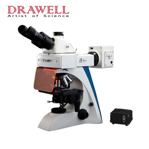
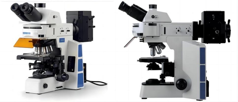
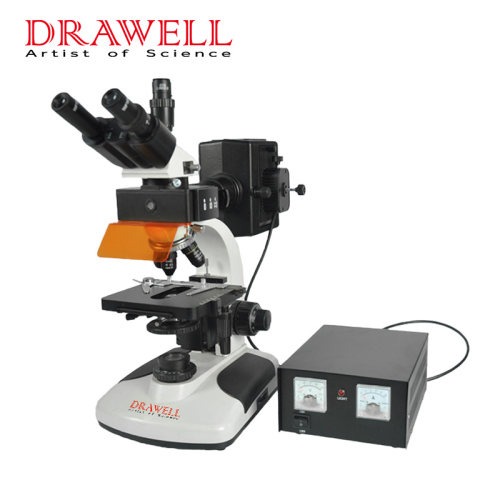
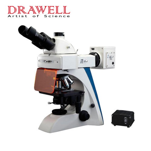
A fluorescence microscope is a strong tool for investigating and observing microscopic structures and processes that are widely utilized in many scientific domains. Its ability to detect and intensify fluorescence, a phenomenon in which certain chemicals emit light when exposed to specific wavelengths of light, makes it a powerful tool in research, diagnostics, and other applications. In this article, we will focus on the topic that when we use a fluorescence microscope, exploring the various applications of fluorescence microscopes and the valuable insights they offer to researchers and scientists across different disciplines.

Fluorescence Microscope Used for Cellular and Molecular Biology
The fluorescence microscope has transformed cellular and molecular biology. Scientists can visualize individual molecules or structures within cells with extreme precision by using fluorescent dyes or proteins. Fluorescence microscopes provide critical insights into the complex realm of cellular dynamics, from analyzing subcellular organelles like the nucleus, mitochondria, and endoplasmic reticulum to tracking cellular processes like protein synthesis, mitosis, and apoptosis.
Fluorescence Microscope Used for Immunology and Pathology
In immunology and pathology, a fluorescence microscope plays a key role in visualizing and studying cellular responses to infections and diseases. Immunofluorescence techniques help identify and localize specific antigens in tissues, allowing researchers and pathologists to diagnose and understand various diseases, including autoimmune disorders, cancer, and infectious diseases. Fluorescently labeled antibodies and probes facilitate the detection of target molecules, offering a detailed understanding of cellular interactions and immune responses.

Fluorescence Microscope Used for Neuroscience
Fluorescence microscopy is important in immunology and pathology for seeing and analyzing cellular responses to infections and illnesses. Immunofluorescence techniques aid in the identification and localization of specific antigens in tissues, helping researchers and pathologists to diagnose and comprehend a wide range of diseases such as autoimmune disorders, cancer, and infectious diseases. Fluorescently labeled antibodies and probes make it easier to detect target molecules, allowing for a more in-depth understanding of cellular interactions and immune responses.
Fluorescence Microscope Used for Live Cell Imaging
Another field that benefits immensely from fluorescence microscopy is neuroscience. This technique is used by researchers to investigate the brain’s complicated neural networks and synaptic connections. Scientists can watch neural activity, analyze neural development, and investigate the mechanisms underlying brain illnesses such as Alzheimer’s and Parkinson’s disease by labeling specific neurons or neural structures with fluorescent markers.
Fluorescence Microscope Used for Genetics and Molecular Diagnostics
Fluorescence in situ hybridization (FISH) is a technique for detecting and visualizing specific DNA sequences within cells or tissues using fluorescence microscopy. FISH has evolved into an important tool in genetics and molecular diagnostics, enabling for the detection of genetic disorders, chromosomal rearrangements, and gene expression patterns. Prenatal testing, cancer diagnosis, and genetic studies all make extensive use of it.
Fluorescence Microscope Used for Environmental and Material Sciences
Fluorescence microscopy is used in environmental and material sciences to examine the composition and properties of materials, as well as to assess environmental samples. Fluorescent labels can be used by researchers to analyze the distribution of certain molecules in materials or to detect pollutants and contaminants in the environment. This allows for the microscopic characterization of materials and aids in environmental monitoring and pollution management.

Fluorescence Microscope Used for Microbiology and Virology
Fluorescence microscopy is a significant tool in microbiology and virology for viewing and analyzing bacteria and viruses. It enables scientists to study the appearance and behavior of bacteria, fungi, and viruses, allowing them to conduct research on pathogenic mechanisms and antimicrobial medicines. Fluorescence-based assays are also utilized to detect and quantify microbial populations in environmental or clinical samples.
Summary
The fluorescence microscope has changed the face of modern science, giving researchers a formidable tool for investigating and comprehending the microscopic world. Its applications range from cellular and molecular biology to immunology, neuroscience, genetics, and environmental sciences, among others. Fluorescence microscopy is at the vanguard of scientific inquiry, yielding revolutionary discoveries, thanks to constant developments in fluorescent dyes, probes, and imaging techniques.


