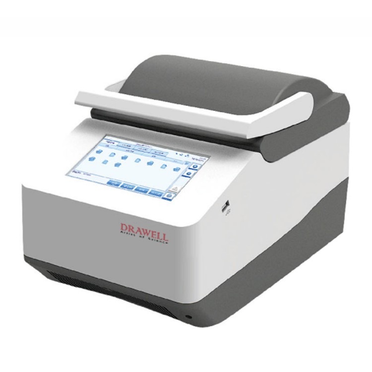
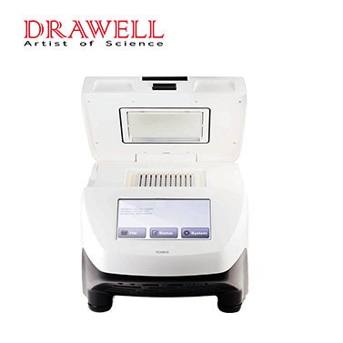
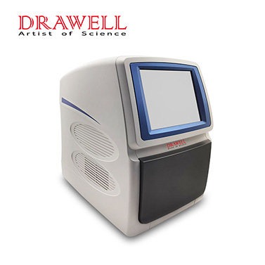
This article will explain to you how to use Polymerase chain reaction (PCR) to measure immune proteins through specific experimental procedures.
Materials and Methods
1. Preparation of labeled specific antibody
There are glycosyl groups on the Fc fragment of immunoglobulins (such as IgG, IgM), so Biotin-hydrazide, which can combine with glycosyl groups, can be used for * labeling.
(1) The purified antibody (IgM or IgG, 0.1~1.0ml) was dialyzed in labeling buffer (0.1Mol/L NaAc, pH 5.5, 0.1Mol/L NaCl) at 4°C for one night.
(2) Pipet 0.5 ml into a microcentrifuge tube, add sodium peroxygen solution to a final concentration of 10 mMol/L, and incubate on an ice bath for 30 min in the dark to oxidize the glycosyl groups on the antibody molecule.
(3) The oxidized antibody was passed through a Sephadex G25 PD-10 prepacked column equilibrated with PBS to separate it from the sodium peroxy. Collect protein peaks.
(4) Add Biotin-LC-hydrazide (Pierce) to the antibody tube to a final concentration of 5 mMol/L, and incubate for 1 h at room temperature on a shaker.
(5) Equilibrate the Sephadex G25 prepacked column with PBS containing 0.02% NaN3, and separate the *-labeled antibody molecules from the free *. The protein peaks were collected and stored at -20°C.
2. Preparation of Labeled DNA Fragments
As the reporter DNA fragment is to be coupled with the antibody, it should be ensured that there is no homologous sequence in the body DNA from which the antigen to be tested is derived. For example, the sequence of Escherichia coli can be used as the reporter DNA to detect human-derived specimens. The size of the DNA fragment is 300~500bp, and the * mark can be done by PCR method. A pair of primers are synthesized according to the nucleotide sequence of the reporter DNA, and one of the primers has a * mark on the 5′-end base.
Mix the following reagents according to the table in a 0.5ml PCR tube.
| PCR System Composition | ||
| Reagent | Added Amount | Final Concentration |
| 10x PCR buffer | 10.0ul | 1× |
| 2.5mM 4dNTP mix | 8.0ul | 0.2mM |
| 50uM* labeled upstream primer | 1.0ul | 0.5uM |
| 50uM* labeled downstream primer | 1.0ul | 0.5uM |
| 15mM template DNA | 1.0ul | 1.0ug |
| Taq DNA polymerase | 1.0ul | 5U |
Bring the ingredients to the table above 100uL with water. It is then covered with liquid paraffin after mixing.
Heating at 95°C for 5min, and then amplified on the PCR machine according to the following procedure: 94°C for 1min; 55°C for 1min; 72°C for 1min; 40 cycles. Finally, an equal amount of phenol-chloroform was added for extraction, then 10ul of 3Mol/L Kac and 250uL of absolute ethanol was added, and the mixture was placed at -70°C for 30min.
The DNA fragments were pelleted by centrifugation and washed once with 70% ethanol. The precipitated DNA was dried, dissolved in TE buffer, and stored at -20°C.
3. Preparation of Antibody-Avidin-DNA Complexes
Mix the *-labeled antibody and avidin in PBS containing 1 mg/ml BSA at an equal molecular concentration, incubate for 30 min at room temperature, then add the *-labeled DNA fragment at twice the molecular concentration, continue to incubate for 30 min, and then add 10 times the molecular concentration. *, saturates the binding site on the avidin molecule. After passing through a gel filtration column, the complex was separated from the unbound monomer, BSA was added to reach 1 mg/ml, and it was frozen at -20°C after aliquoting.

4. Immuno-PCR
(1) According to the conventional ELISA method, dilute the antigen with saturation buffer, add it to a 96-well plastic plate or 0.5ml PCR tube, and place it at 4°C overnight.
(2) Wash three times with PBS, then add 200ul blocking solution (PBS contains 10mg/ml BSA, 1mg/ml protamine DNA) to each well, and incubate at room temperature for 30min.
(3) Wash three times with TETBS (its formula: 20mMol/L EDTA, 0.02% NaN3).
(4) Dilute the antibody-avidin-DNA complex in TETBS containing 1mg/ml BSA and 0.1mg/ml protamine DNA, add 50ul to each well, and incubate at room temperature for 1h.
(5) Wash 5 times with TETBS, then place the plastic plate or tube upside down on absorbent paper and tap to control the moisture.
(6) Add 50ul PCR reaction solution to each tube.
| Reaction Liquid Composition Table | ||
| Reagent | Added Amount | Final Concentration |
| 10x PCR buffer | 5.0μl | 1× |
| 2.5mM 4dNTP mix | 4.0μl | 0.2mM |
| 50uM* labeled upstream primer | 0.5μl | 0.5uM |
| 50uM* labeled downstream primer | 0.5μl | 0.5uM |
The reaction solution was added with water to 50 μL, then mixed well and covered with liquid paraffin.
Heated at 95°C for 5min, and then amplified for 35 cycles on the PCR instrument according to the following procedure: 94°C for 1min; 55°C for 1min; 72°C for 1min. After extension at 72°C for 10min.
Take 5ul of each tube for agarose or polyacrylamide gel electrophoresis, and observe the results after staining with ethidium bromide. If a radionuclide label is added during PCR, X-ray imaging can be used. You can also do Southern blot after gel electrophoresis and hybridize with specific probes to further improve its specificity and sensitivity.
Matters Needing Attention
A critical step in this experiment is obtaining the appropriate antibody-DNA complexes. The method of conjugating *-labeled antibody to *-labeled DNA with streptavidin because each streptavidin molecule can bind to four * molecules, the reaction conditions should be optimized so that each streptavidin molecule can bind to four * molecules Molecules can bind both antibody molecules and DNA fragments.
In addition, chemical methods can also be used to covalently couple DNA fragments to antibody molecules, that is, antibody molecules and DNA fragments modified with amino acids at the 5′ end are activated with different bifunctional coupling agents, and then coupled together through spontaneous reactions, For example, N-Succinimidyl-S-acetyl thioacetate (SATA) is used to activate amino-modified DNA fragments, Sulfo-Succinimidyl4-(maleimidomethyl) Cyclohexane-1-Carboxylate (Sulfo-SMCC) is used to modify antibody molecules, and then the two are combined in a small tube. The two were coupled together by adding Hydroxylamine hydrochloride.
PCR (Polymerase chain reaction) is highly sensitive. Therefore, any non-specific binding of antibodies to labeled DNA can lead to serious background problems. Therefore, it is necessary to wash as thoroughly as possible after adding the antibody and labeling the DNA.
Even if some specifically bound antibody or labeled DNA is washed away, it can be compensated by increasing the number of PCR cycles later. In addition, the use of effective blocking agents is also very important to prevent non-specific binding.
We can use skimmed milk powder and bovine serum albumin as a protein-blocking agent, and protamine DNA as a nucleic acid-blocking agent. Another important factor in preventing background signals is contamination control, which is a problem with all sensitive detection systems. Even if every step of the experiment is done carefully, repeated use of the same primers and labeled DNA can produce false positive signals.
An advantage of PCR (Polymerase chain reaction) is that the marker DNA sequence* is artificially selected. Thus the labeled DNA and its primers can be changed frequently to avoid false positive signals due to contamination.
PCR (Polymerase chain reaction) can detect samples that cannot be detected by conventional immunological methods. Therefore, the application of immune PCR can detect antigens at the microscopic level (single cell), and quantitative PCR products can estimate the number of antigens in a sample.

