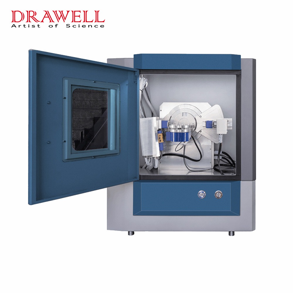
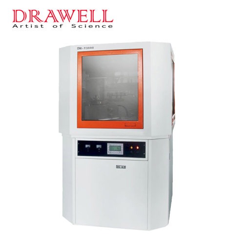
Imagine a tool that can peer into the very fabric of materials, revealing their hidden secrets about atomic arrangement, crystallinity, and even strain. This powerful instrument exists in the form of the X-ray diffractometer, a marvel of scientific ingenuity that unlocks the mysteries of the microscopic world. In this article, we’ll embark on a journey to understand the X-ray diffractometer, exploring its working principles, safety precautions, diverse applications, and concluding with some valuable resources for further exploration.
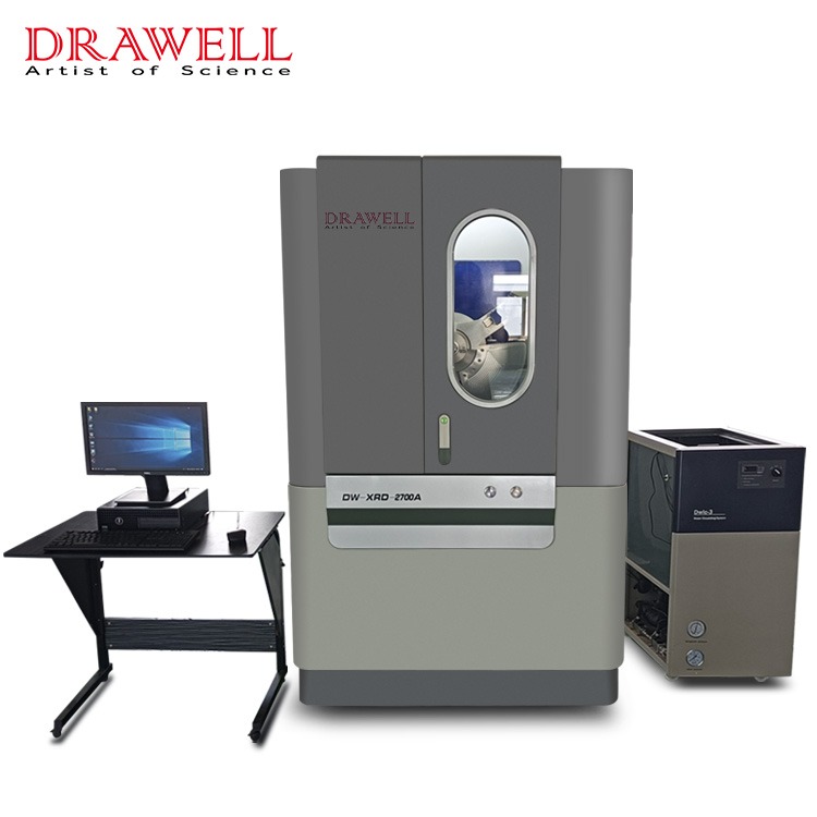
Working Principle of the X-Ray Diffractometer
The X-ray diffractometer isn’t just a box that spits out information about materials; it’s a sophisticated dance between X-rays and atoms, a tango of waves and matter revealing the intricate choreography of nature at the smallest scales. To fully appreciate its power, let’s dissect this dance move by move
1. The Stage is Set
The X-ray source: Imagine a high-voltage ballet, where electrons pirouette under extreme acceleration, eventually slamming into a metal target. This collision unleashes a burst of electromagnetic energy – X-rays, shorter and more energetic than visible light.
The Sample: Our spotlight focuses on the sample, a tiny dancer poised to reveal its secrets. Whether it’s a crystalline powder, a single, gleaming gem, or a thin film hoping to be noticed, the sample stands ready to interact with the X-ray spotlight.
2. The Dance Begins
The Encounter: The X-rays, like energetic photons, waltz onto the stage, bathing the sample in their invisible glow. As these photons brush past the atoms in the sample, a fascinating phenomenon occurs: the atoms act as tiny mirrors, scattering the X-rays in specific directions.
The Bragg Condition: But not all scattered photons make it to the audience (the detector). Only those that fulfill a specific rule, the Bragg condition, get a standing ovation. This rule dictates that the scattered X-rays must be “in step” with each other, their waves reinforcing each other like synchronized swimmers. This “in-step” condition depends on the spacing between the atoms in the sample’s crystal lattice, like the distance between two dancers performing a coordinated routine.
3. The Diffraction Pattern Emerges
The Ripples on the Pond: Imagine throwing a stone in a still pond; ripples emerge, spreading outwards. Similarly, the scattered X-rays that meet the Bragg condition create a “ripple pattern” on the detector. This pattern, unique to the sample’s internal structure, is like a fingerprint, holding the key to its secrets.
4. Interpreting the Dance
Decoding the Ripples: Scientists, the skilled choreographers, analyze the diffraction pattern, measuring the angles and intensities of the “ripples.” By deciphering this code, they can:
- Measure the unit cell parameters: These define the basic building block of the crystal lattice, like the dimensions of the stage on which the atoms dance.
- Identify the space group: This describes the symmetry of the crystal arrangement, like the specific ballet sequence being performed.
- Uncover the presence of different phases: If multiple dances are happening within the sample, the diffraction pattern reveals their presence, like identifying additional dancers on the stage.
- Gauge the crystallite size and strain: These tell us about the size and imperfections of the crystals, like the precision and grace of the dancers’ movements.
Precautions for Using the X-Ray Diffraction Machine
While the X-ray diffractometer unlocks secrets about the material world, its own operation demands careful attention to safety. X-rays, despite their invaluable analytical power, are ionizing radiation capable of damaging human tissue. Therefore, safeguarding oneself during operation is paramount. Here’s a deeper dive into the essential safety precautions:
Shielding Yourself
- Lead Aprons and Gloves: The first line of defense is personal protective equipment (PPE). Lead-lined aprons and gloves, specifically designed to absorb X-rays, shield vulnerable organs and extremities. The lead thickness should be appropriate for the X-ray source and typical exposure times.
- Safety Glasses: Protecting the eyes is crucial, as direct X-ray exposure can harm the retina. Lead-lined glasses with side shields offer comprehensive protection without impeding vision during operation.
- Footwear and Lab Coats: While the primary focus is on shielding vital areas, wearing closed-toe shoes and a lab coat serves as an additional barrier against accidental exposure to scattered radiation.
Operational Protocols
- Training and Authorization: Only trained and authorized personnel should operate the X-ray diffractometer. This ensures familiarity with safety protocols, operating procedures, and emergency response plans.
- Interlocks and Warning Lights: The diffractometer should be equipped with interlocks that automatically shut down the X-ray source if safety barriers are breached. Always heed warning lights and alarms, indicating potential hazards or operational errors.
- Sample Preparation and Handling: Specific procedures for sample preparation and handling minimize exposure risks. This includes using appropriate tools, handling samples within shielded enclosures, and ensuring proper containment of potentially hazardous materials.
- Radiation Monitoring and Dosimetry: Monitoring the lab environment and wearing personal dosimeters is crucial to track individual radiation exposure and ensure safe operating limits are not exceeded.
Maintaining the Machine
- Regular Maintenance and Calibration: Proper maintenance and calibration of the diffractometer ensure optimal performance and safety. Regular checks for leaks, malfunctioning interlocks, and worn-out components are essential. Calibration ensures accurate measurements and minimizes the need for extended exposure times, further reducing radiation risks.
- Emergency Procedures: Familiarity with emergency procedures for accidental exposure, equipment malfunction, or fire is vital. Having a clear plan and readily available safety equipment like lead blankets and emergency eyewash stations minimizes risks and ensures prompt response in case of an incident.
Types of X-Ray Diffraction Machines and their Applications
The X-ray diffractometer, like a versatile chameleon, takes on different forms to meet the unique needs of various scientific disciplines. Here, we’ll shed light on three prominent types and delve into the specific applications they excel in:
1. Powder Diffractometers: The Workhorses of Material Identification
Imagine a detective meticulously analyzing fingerprints to identify a suspect. Powder diffractometers operate in a similar way, albeit with X-rays and crystalline powders. These workhorses of material characterization utilize a broad beam of X-rays to bombard a powdered sample. As the rays scatter off the countless crystallites within, a characteristic fingerprint-like pattern emerges on the detector. By analyzing this pattern, scientists can:
- Identify unknown materials: By comparing the obtained pattern to a vast database of known crystal structures, scientists can pinpoint the exact identity of the mystery powder, be it a mineral, pharmaceutical compound, or a newly synthesized material.
- Quantify different phases: If a sample contains multiple crystalline phases, the diffractometer reveals their relative abundance, providing insights into the material’s composition and potential behavior.
- Track phase transitions: Studying the changes in the diffraction pattern under varying conditions, like temperature or pressure, allows scientists to monitor and understand phase transitions, such as a material changing from ice to water or vice versa.
2. Single-Crystal Diffractometers: Unveiling the Atomic Ballet
While powder diffractometers provide a holistic view of the crystalline landscape, single-crystal diffractometers offer a high-resolution glimpse into the intricate waltz of atoms within a single crystal. By directing a narrow beam at a precisely oriented crystal, this type of diffractometer generates a detailed map of the atomic positions, revealing:
- Crystal structure determination: This is the Holy Grail of crystallography, where the precise arrangement of atoms within the unit cell is mapped, offering crucial information for understanding a material’s properties and behavior.
- Bond lengths and angles: The diffractometer precisely measures the distances and angles between atoms, providing insights into the nature of chemical bonds and the forces governing the crystal structure.
- Molecular orientation and conformation: In complex molecules like proteins, single-crystal diffractometers reveal the intricate folding patterns and orientations of atoms, vital for understanding their function and designing drugs.
3. X-ray Reflectivity Diffractometers: Probing the Surface Secrets
While the first two types delve into the bulk of materials, X-ray reflectivity diffractometers shift their focus to the tantalizing world of surfaces and interfaces. Imagine shining a grazing light across a rippled lake and observing the reflections. Similarly, this type of diffractometer utilizes a low-angle X-ray beam that gently skims across thin films and interfaces, revealing their hidden secrets:
- Film thickness and roughness: By analyzing the intensity and pattern of the reflected X-rays, scientists can precisely measure the thickness of thin films, from a few nanometers to several micrometers, and even assess their surface roughness.
- Layered structures: For multi-layered thin films, like those used in solar cells or electronics, reflectivity diffractometers unveil the composition and thickness of each layer, essential for optimizing their performance.
- Interface characterization: Studying the reflectivity pattern at the interface between two materials provides insights into their bonding, strain, and other interfacial properties, crucial for understanding phenomena like charge transfer or friction.
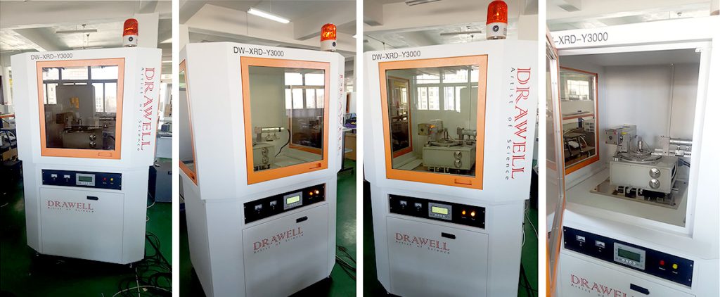
Summary
The X-ray diffractometer stands as a testament to human ingenuity, offering a powerful window into the hidden world of materials. Its ability to unravel the secrets of crystal structures and provide valuable information about a material’s composition, phase, and physical properties makes it an indispensable tool in diverse scientific fields. As research and technology advance, X-ray diffraction techniques continue to evolve, promising even deeper insights into the microscopic realm with ever-increasing precision and sensitivity.
Recommended Books
“Elements of X-ray Diffraction” by B.D. Cullity and S.R. Stock
“The Powder Diffraction File: A Reference Intensity Database for Identification of Crystalline Materials” by International Centre for Diffraction Data
“Introduction to X-ray Crystallography” by William Clegg

